Full Body MRI: What It Can Show and What It Cannot
A full body MRI can examine multiple regions of the body in one imaging session.
That sounds comprehensive, but comprehensive does not mean complete.
MRI can provide detailed structural information without using ionizing radiation. A whole body MRI may reveal findings across several organs, soft tissues, bones, and other anatomical regions, depending on the protocol used. What it does not do is replace every targeted imaging study, laboratory test, or recommended screening program.
That distinction matters.
For physicians, the value of a full body MRI is not simply the number of structures captured in the scan. Its usefulness depends on why the imaging was performed, the patient's medical and family history, existing risk factors, previous studies, and what happens after a finding is reported.
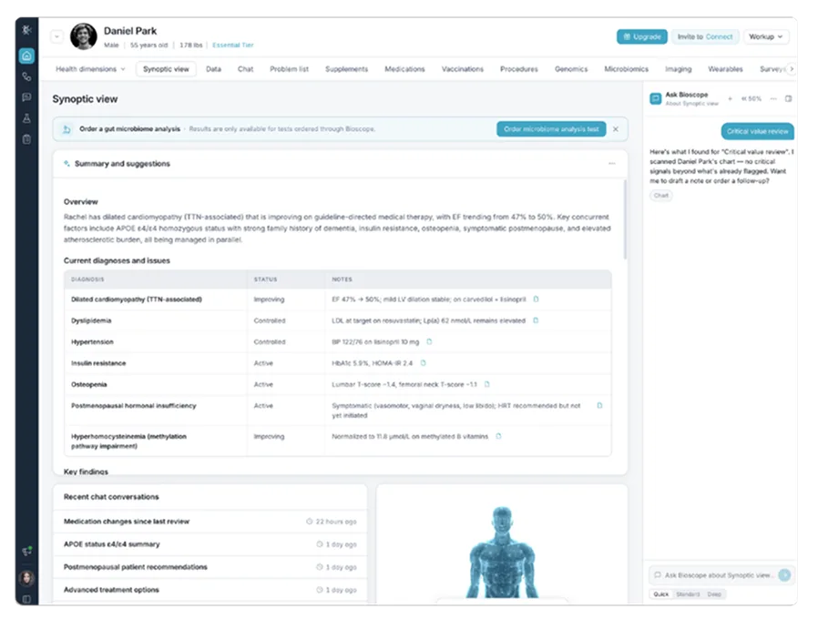
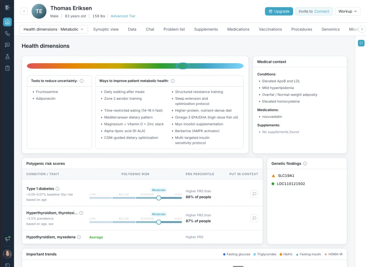
Bioscope.ai already includes whole-body MRI organ analysis within its imaging data. The platform's broader approach is to place imaging beside other forms of patient information rather than treating the scan as a complete health assessment on its own.
What Is a Full Body MRI?
MRI uses a strong magnetic field and radiofrequency signals to produce detailed images of structures inside the body.
A full body MRI, also called a whole body MRI, extends this approach across multiple anatomical regions instead of concentrating on one specific area.
The exact coverage depends on the protocol.
Whole-body MRI protocols can include the head, neck, chest, abdomen, pelvis, bones, and limbs. Different MRI sequences are used to characterize anatomy and identify findings that may require closer assessment.
This is different from a targeted MRI.
If a physician needs a detailed examination of a specific joint, organ, or neurological concern, a dedicated MRI protocol can be optimized for that clinical question. A broader scan covers more territory but may not provide the same level of detail for every structure.
That trade-off is important when interpreting the results.
Full Body MRI and Targeted MRI Answer Different Questions
A targeted MRI starts with a defined problem. A patient may have neurological symptoms, a musculoskeletal injury, an abnormal laboratory finding, or another reason to examine one area in detail. The imaging protocol is then designed around that question.
A full body MRI starts from a wider field of view.
It may identify abnormalities in areas that were not the original focus of the examination. That broader coverage can be useful, but it also changes the type of decisions that follow.
More anatomy means more opportunities to find something unexpected.
Some of those findings may be important. Others may be benign variations or abnormalities that do not affect the patient's health.
The physician still needs to decide which findings deserve action.
What Can a Full Body MRI Show?
Whole-body MRI provides structural information.
Depending on the protocol, it can show abnormalities involving soft tissues, organs, bones, and other anatomical structures across several regions.
In some higher-risk populations, whole-body MRI also has established roles in cancer surveillance.
That does not mean the same recommendation applies to every healthy person.
The meaning of a finding depends heavily on who is being scanned and why.
An abnormality identified in someone with a known genetic cancer predisposition may require a different response from a similar finding in an asymptomatic person with no known risk factors.
This is why patient history cannot be separated from imaging interpretation.
A Wider Scan Also Finds Incidental Findings
One of the most important concepts in whole-body imaging is the incidental finding.
An incidental finding is something identified on imaging that was not the original reason for performing the examination.
Some incidental findings turn out to be clinically important.
Many do not.
The challenge is distinguishing between the two.
Broad imaging can reveal nonspecific abnormalities that lead to additional testing, follow-up imaging, procedures, cost, and patient concern without necessarily changing long-term outcomes.
That does not make incidental findings unimportant.
It means they need proportionate follow-up.
A Finding Is Not the Same as a Diagnosis
MRI produces images. It does not independently determine what every abnormality means.
A radiologist may identify a lesion, structural variation, signal abnormality, or another finding that requires further interpretation.
Sometimes the imaging characteristics strongly support a benign explanation.
Sometimes further evaluation is appropriate.
And sometimes the answer remains uncertain until the finding is compared with prior imaging or examined with a more targeted study.
This is why language matters.
A full body MRI should not be described as a test that simply “finds hidden disease.”
What it does is identify imaging findings.
The clinical team then determines whether those findings are expected, incidental, potentially significant, or in need of additional investigation.
Bring imaging into the full patient record
See how longevity medicine software places MRI findings beside labs, genomics, wearables, and history for physician review.
Schedule a DemoRisk Changes the Meaning of the Scan
The same image can carry different implications in different patients.
A patient's personal history, family history, genetic risk, previous diagnoses, symptoms, and prior imaging all affect how a new finding should be interpreted.
A scan performed in a higher-risk individual is not interpreted the same way as a scan performed in an asymptomatic person from the general population.
The likelihood that a finding represents significant disease changes with the patient's background.
This is one reason a full body MRI report cannot be interpreted well without clinical information.
The scan shows anatomy.
The patient's history changes what that anatomy means.
Full Body MRI Does Not Replace Standard Screening
This is one of the most important limitations to understand.
Whole-body MRI should not be treated as a substitute for every established screening test.
Certain organs and diseases are better evaluated with dedicated screening methods or targeted examinations.
A broader scan does not automatically mean better screening.
The right test depends on the clinical question.
Mammography, colorectal screening, cervical screening, targeted CT, ultrasound, endoscopy, or another test may still be more appropriate depending on the patient and the condition being assessed.
Full body MRI should therefore be considered one imaging option, not a universal replacement for established screening pathways.
Interpret findings in context
Explore precision medicine software for physicians that connects imaging with prior scans, risk factors, and other patient data.
Schedule a Personalized DemoWhat a Full Body MRI May Miss
No imaging test sees everything.
Whole-body MRI has limitations related to the structures being examined, the sequences used, movement during the scan, technical quality, lesion size, and the type of abnormality being investigated.
Some areas require dedicated imaging.
Small abnormalities may be better characterized with targeted MRI sequences, CT, ultrasound, mammography, endoscopy, or another modality, depending on the clinical question.
Whole-body protocols also have recognized blind spots.
That means a normal full body MRI should not be interpreted as proof that no disease is present.
It is one source of clinical information. A physician still needs to consider symptoms, risk, laboratory testing, recommended screening, physical examination, and other evidence.
Previous Imaging Can Change the Interpretation
A new scan is often more useful when there is something to compare it with.
If an abnormality was already present on a previous study and has remained stable, that history can affect how it is interpreted.
If a finding is new or has changed, it may deserve closer attention.
Comparison also helps avoid unnecessary workups.
Without previous imaging, a radiologist may have less information about whether a finding represents a long-standing benign feature or a recent change.
This makes imaging history part of the clinical record, not simply an archive of old scans.
For longevity-focused care, that becomes particularly relevant when patients undergo testing at different points in time.
Where Full Body MRI Fits in Longevity Medicine
Longevity medicine often looks at more than one moment in a patient's health.
Physicians may review laboratory trends, body composition, cardiovascular measures, genomics, wearables, imaging, medical history, and other information across time.
Full body MRI can add structural information to that process.
That makes it relevant to longevity medicine software, where imaging can sit beside other health data rather than being treated as a separate endpoint.
The scan should not become a longevity score.
It should not be used to claim that a patient is healthy simply because no significant abnormality was identified.
Its role is narrower and more useful than that: MRI can add another layer of information that the physician can interpret together with the rest of the patient's record.
Structural Imaging and Biological Data Answer Different Questions
A scan, a blood test, a genome, and a wearable do not measure the same thing.
That is exactly why combining them can be useful.
MRI provides anatomical information.
Laboratory testing measures biochemical and physiological markers.
Genomics can provide information about inherited variation.
Wearables capture selected signals during daily life.
Medical history adds events, diagnoses, medications, symptoms, and previous clinical decisions.
None of these sources should be expected to replace the others.
Within precision medicine software for physicians, the goal is to interpret different forms of information in relation to the individual patient.
Whole-body MRI contributes imaging evidence to that process.
Why More Data Is Not Automatically Better
Modern medicine can generate extraordinary amounts of information.
The challenge is deciding what deserves attention.
Whole-body imaging illustrates this particularly well. A single scan can generate findings across many anatomical regions.
If every abnormality is treated with the same urgency, patients can be exposed to unnecessary follow-up.
If everything is dismissed, meaningful findings could be missed.
The answer is not simply to collect more data.
It is to interpret data according to clinical significance.
That requires a physician to understand the patient's risk profile, symptoms, previous results, and reason for testing.
The same principle applies beyond MRI.
More laboratory results, more wearable measurements, or more genomic information do not automatically produce better care. Clinical value comes from knowing what matters.
How Bioscope.ai Places Imaging in a Wider Patient View
Bioscope.ai does not treat imaging as the only source of truth about a patient.
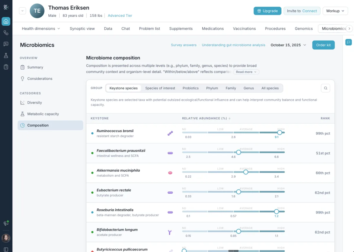
Its platform brings imaging into a wider clinical model that also includes whole-genome data, medical records and laboratory results, wearables, microbiome information, and information gathered during the physician visit.
Whole-body MRI organ analysis fits within that imaging layer.
This approach is particularly relevant when a scan produces a finding that cannot be understood from imaging alone.
The physician may need to see whether laboratory results support the same concern, whether there is a relevant family history, whether genomic information changes risk, or whether previous imaging shows that the finding has been stable.
That is where integration becomes more useful than simply displaying another scan result.
The Best Question Is Not “Did the Scan Find Something?”
A full body MRI can produce a long report.
The length of that report does not tell us how healthy or unhealthy the patient is.
A better set of questions is whether the finding matters for this patient, whether it fits the patient's history or risk profile, whether it has changed, and whether it requires targeted follow-up.
These are interpretation questions, not imaging questions.
They are also why physician involvement remains essential when broad imaging is added to a longevity or precision medicine program.
A Full Body MRI Is One Layer of the Health Record
Full body MRI can provide a broad structural view across multiple regions of the body.
That is valuable information.
But the scan does not eliminate uncertainty, replace standard screening, or provide a complete assessment of health.
Its strongest role is more measured.
A whole body MRI adds imaging evidence that can be interpreted in relation to the patient's medical history, family history, risk factors, prior scans, laboratory results, genomics, and other relevant information.
For Bioscope.ai, this fits naturally with a physician-led approach to complex patient data.
The goal is not to make one scan answer every question.
It is to give physicians enough connected information to decide which findings matter, which need follow-up, and which should remain what they are: findings rather than diagnoses.
See Bioscope.ai in Action
Discover how Bioscope.ai connects imaging, genomics, labs, and wearables so physicians can interpret findings in the context of each patient.